The visual exam is the third part of the three-part voice examination. Laryngoscopy and stroboscopy confirm what the history and listening exam predicted — and reveal the structural detail needed to plan treatment.
Tips for Great Laryngoscopy
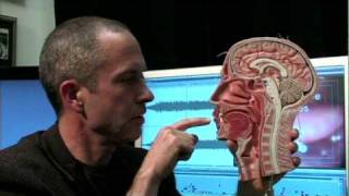
◆ Laryngoscopy
Rigid, flexible, and chip-tip endoscopes each provide a different view; none replaces the others. The recording device matters more than the scope. A complete exam sequence — from equipment selection through stroboscopy — takes 45 to 60 minutes and produces a permanent record.
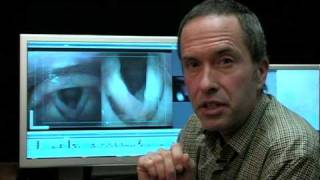
◆ High Definition Techniques
Snapshot examination finds large lesions; high-definition examination finds the precise margins, the capillary pattern, and the mucosal wave that determine diagnosis and surgical planning. Stroboscopy technique and selective color imaging (NBI and iScan) are explained in detail.
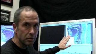
◆ Topical Anesthesia
Flexible chip endoscopy through an anesthetized nose allows prolonged, close examination without the patient’s gag reflex. The 4% lidocaine protocol, Abraham cannula technique, laryngeal gargle, and transcricothyroid injection each serve a specific purpose in the sequence.